
The month of October is often associated with Halloween and people think "hocus pocus" but this time we are promoting point of care ultrasound (POCUS) with a month-long discussion of subjects regarding bedside ultrasound. This is all in conjunction with Practical POCUS which will be having courses at the end of this year. Our first topic, ultrasound for the diagnosis of urolithiasis.
As mentioned above, Practical POCUS has upcoming courses in Missouri at the end of the year. With a special discount code "TOTALEM" you can get 10% off! Remember to register soon since there is an early bird discount already in place and the limited seating is filling quickly.
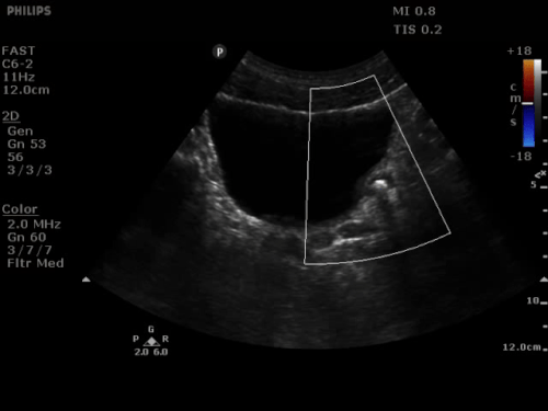
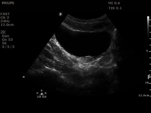
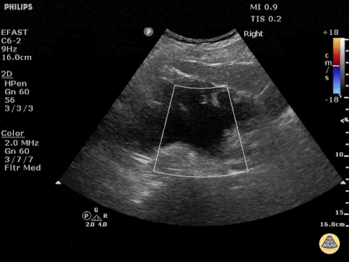
Now to the content you really want. A while back, the American College of Emergency Physicians (ACEP) made a recommendation with the Choosing Wisely campaign to reduce the number of CTs being performed. Essentially, they recommend avoiding CT in young otherwise healthy patients less than 50 years old with a history of known kidney stones that are presenting with symptoms consistent with uncomplicated renal colic. Instead, they recommend follow-up with primary care and/or urology. Kevin Klauer, who has been on the podcast in the past, talked about some of the evidence when it comes to CT versus ultrasound for such patients. With concerns for radiation and its complications on the rise, ultrasound has a potential benefit. However, there are some quick pearls with performing ultrasound alone in such patients. First, usually we do not see stones in the ureter directly. However, as a couple of images below demonstrate, sometimes you can get lucky. Below, you can see two images from The POCUS Atlas, showing one of these stones at the left ureterovesical junction (UVJ) with an observable "twinkle artifact" on the left image. This happens when highly echogenic objects that are stationary create a false sense of movement. The exact cause of why this happens is unclear, but can be helpful in the detection of stones. A long axis view identifies this stone as 7mm in length.
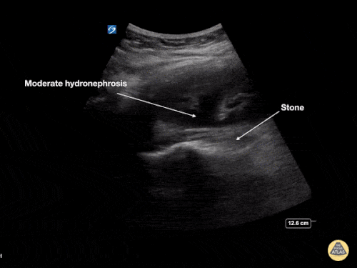
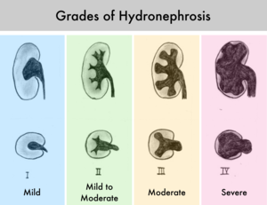
The recent paper we are about to discuss reviews looking for degrees of hydronephrosis and their importance. Mild hydronephrosis will have some fluid in the renal pelvis but little to no involvement of the calyces. Moderate hydronephrosis means that the fluid in the renal pelvis extends into the calyces, as well. Severe hydronephrosis is when there is fluid in the renal pelvis with significant involvement of the calyces along with thinning of the renal parenchyma.
The images here demonstrate all three types along with a graph and comes from The POCUS Atlas. Look closely at the moderate hydronephrosis one to see a 7mm stone that is hyperechoic and causing posterior acoustic shadowing at the ureteropelvic junction (UPJ). Hydroureter can also be seen and can be rather significant.
Before we move on, lets talk about the complications that lurk around the corner. We may not be able to see a stone or there could be multiple stones. We will not usually know this by ultrasound which is all the more reason to ensure follow-up and have shared decision making with patients. More importantly, there are potential high-risk diagnoses that we could miss: abdominal aortic aneurysm (AAA) with rupture, aortic dissection, pyelonephritis, renal infarction, pneumonia, appendicitis, diverticulitis (with abscess), bowel ischemia, bowel perforation, or ovarian torsion.
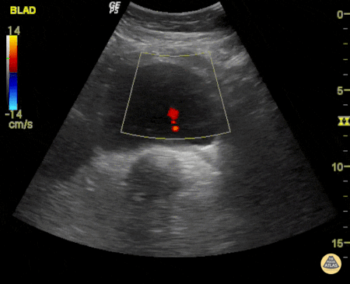
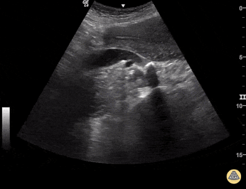
The main one that clinicians see to worry most about is a ruptured AAA or aortic dissection. Some have recommended taking a quick look at the aorta especially those on the older end of the spectrum, have not previously been diagnosed with a stone, or have not had previous/recent assessment of their aorta. One such group is the Ultrasound Podcast which has a Part 1 and Part 2 podcast they did regarding renal ultrasound. If you want to learn more about ultrasound right away, this is a great resource, but still go to a course. One last tip, while you are scanning the bladder look for a ureteric jet. This can be helpful if you are thinking about an obstruction. A good ureteric jet can be reassuring of the lack of an obstruction. If there is enough of a hydroureter, you may see the stone away from the UVJ and UPJ like the one below. Here, you can see a stone and its shadow with the hydroureter again from The POCUS Atlas.
Now on to the recent paper. Academic Emergency Medicine recently published a paper by Wong et al. called The Accuracy and Prognostic Value of Point‐of‐care Ultrasound for Nephrolithiasis in the Emergency Department: A Systematic Review and Meta‐analysis which was also covered in a systematic review snapshot by Annals of Emergency Medicine. In this recent systematic review and meta-analysis, it challenges some of the success with POCUS for the diagnosis. Five studies were included in the analysis for diagnostic accuracy with these ranging from 35.5% to 84.2% compared to a non-contrast CT as the gold standard. The overall sensitivity was 70.2% and the specificity was 75.4% for the 1,773 patients included.
When it came to two of the studies with a total of 1,505 patients that included the degree of hydronephrosis being evaluated, those with moderate to severe hydronephrosis had a sensitivity of 29% but a specificity of 94.4%. This makes a likelihood ratio of 5.22 and a negative likelihood ratio of 0.76 based on this evidence. Hydronephrosis was usually suggestive of a larger stone (>5mm). Although there are limitations, especially with the meta-analysis given the heterogenecity, we should focus on the positives rather than the negatives of a POCUS exam. Patients with moderate or severe hydronephrosis when consistent with the rest of the history, physical, and lab work is highly specific for the presence of a stone in patients with renal colic symptoms. However, patients without hydronephrosis cannot exclude the diagnosis. Let us know what you think by giving us feedback here in the comments section or contacting us on Twitter or Facebook. Remember to look us up on Libsyn and on iTunes. If you have any questions you can also comment below, email at [email protected], or send a message from the page. We hope to talk to everyone again soon. Until then, continue to provide total care everywhere.
1 Comment
d
11/12/2020 11:30:21 am
good Leave a Reply. |
Libsyn and iTunesWe are now on Libsyn and iTunes for your listening pleasure! Archives
August 2022
Categories |
||||||||||
 RSS Feed