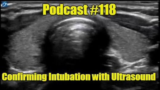
There are many ways to confirm successful intubation. Some are better than others, but they all have limitations. One newer approach is the use of point of care ultrasound (POCUS) to provide real-time confirmation of tube placement. We will talk about how to do this exam and its evidence in this blog and podcast.
Remember, this October is ultrasound month in conjunction with Practical POCUS which has upcoming courses in Missouri at the end of the year. With a special discount code "TOTALEM" you can get 10% off! Remember to register soon since there is limited seating which is filling quickly.
Even quantitative waveform capnography has been shown to be only 65-68% sensitive for detecting the correct endotracheal tube location during cardiac arrest. End-tidal CO2 detection also requires at least five breaths for confirmation which can lead to gastric distention with increased risk for aspiration. Chest x-ray is slow and sometimes difficult to see well. Bedside ultrasound is a valuable alternative especially in cardiac arrest where it can perform much better than quantitative capnography. It also provides immediate feedback when used dynamically. The person using the ultrasound can see in real time esophageal versus tracheal intubation. There are different techniques but the generalized approach is easy to perform. Place the linear ultrasound probe just superior to the suprasternal notch. This should be a transverse view anterior and over the trachea. In patients where you are concerned about needing a possible surgical airway, this is a great time to also look for the cricothyroid membrane in patients where it is not obvious. Check out Cynthia Griffin's post of FOAMfrat for some more details on the airway part of this POCUS exam. Check out the images below for some more detail. Again, The POCUS Atlas is a great source for ultrasound images. In the appropriate placement, you should see a single air filled structure. If this is done dynamically, you can see artifact as the tube is passing through the trachea. However, if you see a second air filled structure, this is evidence of an esophageal intubation. Again, check out the GIF and when doing this dynamically, you can see a second air filled structure suddenly developing which is very concerning for esophageal intubation. If there is any question about cuff placement, moving saline in or out of the cuff creates bubbles which can further assists in identification as shown in the third GIF. Make sure to also assess for lung sliding on both sides. They should be equal (assuming they were prior to intubation) in the amount of movement. Absence of lung sliding should be considering for potential mainstem intubation.
The Annals of Emergency Medicine published an article by Gottlieb, et al. called Ultrasonography for the Confirmation of Endotracheal Tube Intubation: A Systematic Review and Meta-analysis. This well done study incorporated different settings, techniques, and levels of experience.
Although this paper covered adults, the approach is similar for pediatric patients. The Ultrasound Podcast did a great review of one study with a detailed explanation of the approach and some pearls to improve your intubation skills while using POCUS. In the Annals paper listed above, they allowed for a number of techniques to be assessed. We described a dynamic evaluation, but eight of the studies in the paper used dynamic evaluation, nine used static evaluation, and one allowed for either techinque. There were some different ways to confirm placement but most commonly it was the above described technique of looking for two air-filled structures creating an air-mucosa interface (also called the "double tract" sign when referring to esophageal intubation). Three studies did use the presence of fluttering within the trachea as the tube passed through (known as the "snowstorm" sign), and one used a combination of this with conversion of the cricothyroid area from being triangular to round (known as a "bullet" sign). The dynamic approach looking for two air-filled structures is the most straight-forward, but a second person to use the ultrasound machine is not always available. If this is the case, the intubator can then switch roles and check themselves which can still be fairly rapid if the machine is ready and placed appropriately. Alternatively, someone can hold the probe in place while the person intubating can then look up at the screen to further confirm placement. No matter the method, this should be very rapid and if done properly can be faster than any other technique available. In this systematic review and meta-analysis, transtracheal ultrasonography was 98.7% sensitive (95% confidence interval 97.8% to 99.2%) and 97.1% specific (95% confidence interval 92.4% to 99.0%) with a positive likelihood ratio of 34.4 and a negative likelihood ratio of 0.01! The mean time to confirmation was just 13 seconds. The studies had low heterogeneity and the subgroup analyses excellent sensitivity, specificity, and positive and negative likelihood ratios. These studies were also at low risk for bias. 12 of these studies were in the ED but 5 of these studies were in the operating theater. Even with the significant variations, there was no significant difference in the accuracy of these studies. This also holds true for the differing levels of experience. Like many other POCUS exams, this is relatively straightforward and easy to train. Essentially, this is something that we should do more routinely. It is about four times faster than capnography. Even if you do both transtracheal and lung ultrasounds, this only takes 48 seconds based on a previous study. This does not mean you cannot do capnography, but ultrasound is able to additionally identify a potential mainstem which would otherwise have to wait for other means of confirmation such as auscultation (which can be very inaccurate) or chest x-ray (which takes time to set up and perform). In fact, the 2015 advanced cardiac life support (ACLS) guidelines supports the use of POCUS for such an evaluation. Let us know what you think by giving us feedback here in the comments section or contacting us on Twitter or Facebook. Remember to look us up on Libsyn and on iTunes. If you have any questions you can also comment below, email at [email protected], or send a message from the page. We hope to talk to everyone again soon. Until then, continue to provide total care everywhere.
0 Comments
Leave a Reply. |
Libsyn and iTunesWe are now on Libsyn and iTunes for your listening pleasure! Archives
August 2022
Categories |
||||||

 RSS Feed