|
Recently, our own Chip Lange was on The Skeptic's Guide to Emergency Medicine (The SGEM) for Podcast #249 covering ultrasound to confirm endotracheal tube placement. This had previously been discussed in detail on our own Podcast #118 along with the technique. However, after some discussions on social media it was decided to expand on this discussion further regarding the ways to confirm beyond ultrasound.
First, a quick recap. It is vital to not only accurately identify endotracheal tube placement, but to do so quickly. An incorrectly placed tube may not always be identified quickly with traditional techniques. Furthermore, a mainstem intubation carries its own complications including significant desaturation that can lead to cardiac arrest.
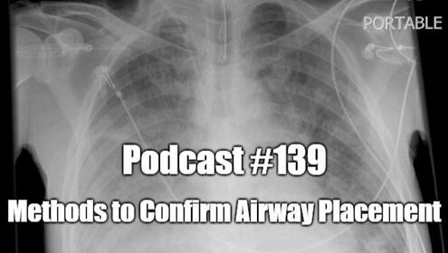
Direct visualization is commonly argued as the best method to confirm placement into the trachea, but anyone who has worked long enough in the field has seen a patient who gets worse despite the person performing the intubation swearing that they saw the tube go through the cords. Does that mean that they are inexperienced or do not know what they are doing? Even the most experienced can make this mistake, especially as the stress increases in a critical situation. Additionally, we may not always be able to directly visualize the vocal cords and must rely on other landmarks. Overall, direct visualization, especially by video where multiple people can confirm placement, is great but it is still not a perfect method. Many people will recognized auscultation is not reliable mainly because lung sounds can be misleading or difficult to hear in a loud environment. We may try to listen to the epigastrium first for air, but it is still a challenge to hear this sound. Additionally, we try to listen for equal lung sounds in order to detect a possible mainstem intubation. In a noisy environment, this is not easily detectable. Pulse oximetry can have a long delay. Critically ill patients can be delayed for approximately a minute. In patients with healthy lungs that need to be intubated for other reasons, mainstem intubation is less likely to cause a significant drop in oxygen saturations at least initially. This ties in with another common technique for traditional confirmation of endotracheal tube placement, the chest x-ray. Although helpful with gauging position and depth, it is generally the slowest technique to confirm placement. It can logistically be challenging to get a portable chest x-ray to the bedside in a timely fashion especially in critical events such as cardiac arrest as this will further delay other lifesaving measures such as high quality CPR. This means this classic "gold standard" is not sufficient in many of our patients. Devices that detect esophageal intubation such as a bulb or alternatively devices that can be attached such as a light to the confirm endotracheal intubation is often slow and requires additional equipment not found in many locations. Additionally, it requires additional steps that only delay fixing the ultimate problem in the intubation attempt. End-tidal carbon dioxide (CO2) detection (continuous waveform capnography, colorimetric, and non-wave form capnography) is one of the preferred methods in intubation to confirm placement. Waveform capnography has the added benefit of being less likely to represent a false positive for esophageal intubation. However, waveform capnography is not as accurate in certain situations such as cardiac arrest. It can also be inaccurate as there are some cases where carbon dioxide may be present in the stomach leading to a false positive especially on color change. This returns to the original point. Point of care ultrasound (POCUS) addresses these problems. It is fast, sensitive, and specific at confirming placement. Especially in dynamic scanning, bedside ultrasound can identify placement as quickly as direct visualization and even faster than end tidal CO2 (ETCO2). Combine assessing the neck and lungs at the same time and the issue with depth is also addressed. Equal lung sliding both before and after endotracheal tube placement reassures that the tube has not been placed in a mainstem fashion. Ultrasound can be done in a variety of environments including those where there is limited access to resources. It can be used on a cramped ambulance, on a loud helicopter, or in a resource poor part of the world that may lack other technologies described above. As discussed on The SGEM, none of these modalities should be used by itself. None work 100% of the time and each is user dependent. With that said, POCUS should be considered as a reasonable method to include in our arsenal for endotracheal tube placement. Let us know what you think by giving us feedback here in the comments section or contacting us on Twitter or Facebook. Remember to look us up on Libsyn and on iTunes. If you have any questions you can also comment below, email at [email protected], or send a message from the page. We hope to talk to everyone again soon. Until then, continue to provide total care everywhere.
0 Comments
Leave a Reply. |
Libsyn and iTunesWe are now on Libsyn and iTunes for your listening pleasure! Archives
August 2022
Categories |
||||||


 RSS Feed